Non Coronary Cusp Echo
Q Tbn 3aand9gctydyi2l5tfld4sx8wzmekvjzopx8sl9l6gk92pt J97smyhg R Usqp Cau

Acute Aortic Regurgitation Circulation
Frontiers Incidental Finding Of An Aorto Right Atrial Fistula In A Patient Undergoing Repair Of A Sinus Of Valsalva Aneurysm Medicine

View Image
Aortic Valve Quiz Echocardiography Ultrasound And Perioperative Medicine
Echocardia Wiki
This quiz will review basic images and normal anatomy of perioperative transesophageal echocardiography.

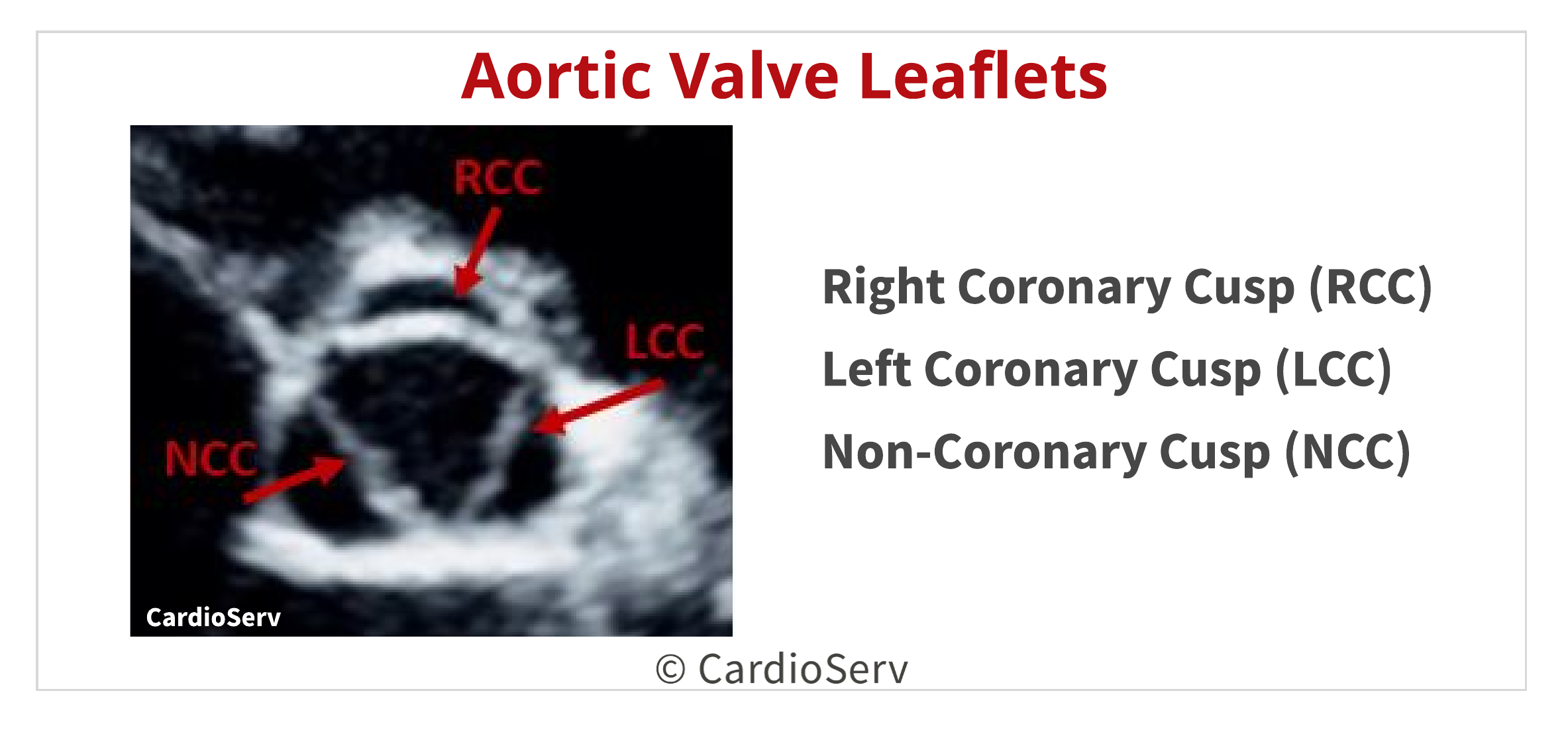
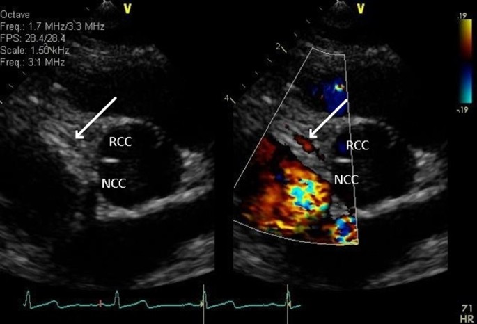
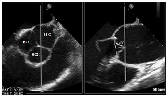
Non coronary cusp echo. Left coronary cusp NCC:. LCC, left coronary cusp;. Right Coronary Cusp (RCC) Left Coronary Cusp (LCC) Non-Coronary Cusp (NCC) Therefore, we can associate the RCC with the right coronary artery and the LCC with the left coronary artery.
Doppler Echocardiography Color Doppler. Isolated thickening of one aortic cusp occurs more commonly in male patients (64%) (p < 0.01) and at an earlier age (65 years) than mitral annular calcification (70 years) (p < 0.001). Each cusp has two free edges, which is shared with the neighboring cusp.
The resolution power of modern day echocardiography is 2mm and the left main ostium is >3.5mm in 99% of population. Blood cultures grew Streptococcus viridance. Non-Coronary Cusp (black arrow).
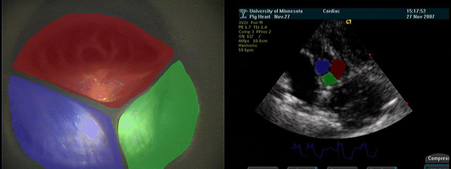
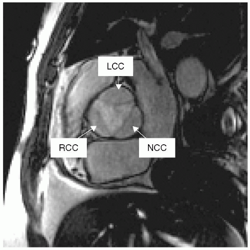
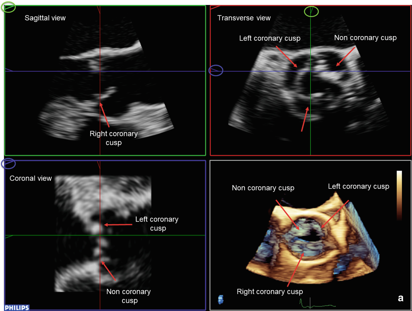
The top, left image shows the short axis view of the aortic valve under direct visualization, with the cusps highlighted in red, green, and blue, respectively. The anterior and posterior leaflets. Apical long axis, non-coronary cusp of the aortic valve ;.
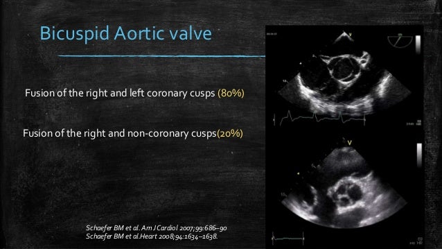
The normal arrangement for the aortic valve is three leaflets or cusps :. A commissure is the space or. Cusp with both coronary arteries arising from the anterior cusp (80% of cases), or fusion of the right and non-coronary cusps resulting in a larger right than left cusp with one coronary artery arising from each cusp (about % of cases).5,6 Fusion of the left and non-coronary cusps is rare.
Unicuspid aortic valves usually have incomplete fusion of all of the leaflets and exhibit aortic stenosis. The heart valves and the chambers are lined with endocardium.Heart valves separate the atria from the ventricles, or the ventricles from a blood vessel.Heart valves are situated around the fibrous rings of the cardiac skeleton.The valves incorporate flaps called leaflets or cusps, similar to a duckbill valve or flutter valve, which are pushed open to allow blood flow and which then close. Log in Sign up.
Diagnosis is most reliable when the two. (closed) and systole (open). Certain coronary anomalies have been associated with sudden cardiac death.
This iPhone, iPod, and iPad Transesophageal Echocardiography Atlas highlights the 28 most common TEE views used in a comprehensive perioperative TEE exam. +3.3) and ascending aorta (4.7cm) (figure 1A and 1B), severe aortic stenosis (aortic. They may be called the left coronary, right coronary and non-coronary cusp.
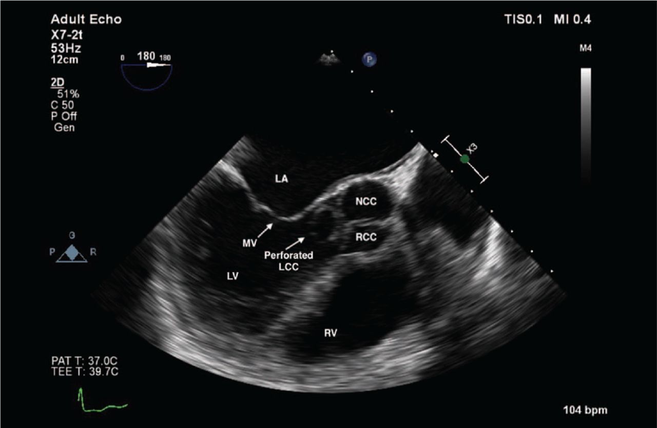
Transthoracic echocardiography revealed normal left ventricular function and a 1.4×1.5-cm mobile mass attached to the aortic aspect of the noncoronary cusp of the aortic valve. ECHO Views is a quick reference tool developed for the beginning to intermediate echocardiographer. The postoperative course was.
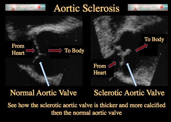
* Aortic sclerosis, although hemodynamically insignificant, it is considered to be as a precursor of aortic stenosis. Echocardiography, Ultrasound, and Perioperative Medicine Moving our Specialties Forward Normal TTE Quiz. Preoperative short-axis images obtained with two-dimensional transesophageal echocardiography, showing a mobile mass attached to the left coronary cusp of the aortic valve and two.
Log in Sign up. If some body says one can’t visualise the coronary artery by echo , it can only reflect their. Apical long axis, left coronary cusp of the aortic valve.
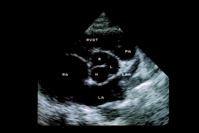
Question 1 - Followup R N L N R L Answer:. This quiz will review basic images and normal anatomy of transthoracic echocardiography. Cusp flail is complete eversion of a cusp into the LVOT (Figure 8A).
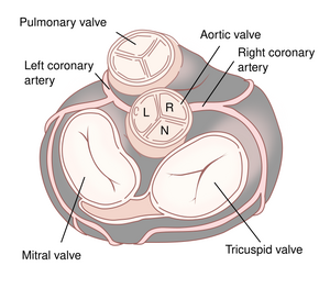
For 146 of the 155 patients, the origin determined by the successful ablation site was at the L-RCC in five, LCC in 13, RCC in six, non-coronary cusp in two, right ventricular outflow tract in 108, left ventricular outflow tract in five, left ventricular epicardium in four, and pulmonary artery in three. The left aortic cusp gives rise to the left coronary artery, the right aortic cusp gives rise to the right coronary artery and the non-coronary cusp does not give rise to an artery. In the parasternal long-axis view the right coronary cusp appears _____ to the non-coronary cusp.
RCC = right coronary cusp;. Content from this web site may not be used or reproduced for non-personal or. He had previously undergone aortic arch replacement and valvuloplasty of a non-coronary cusp with a patch to correct aortic dissection and moderate aortic valve regurgitation through a tear in the non-coronary cusp.
STJ = sino-tubular junction;. Non-atherosclerotic coronary artery disease and sudden death in the young. AOV left coronary cusp 5.
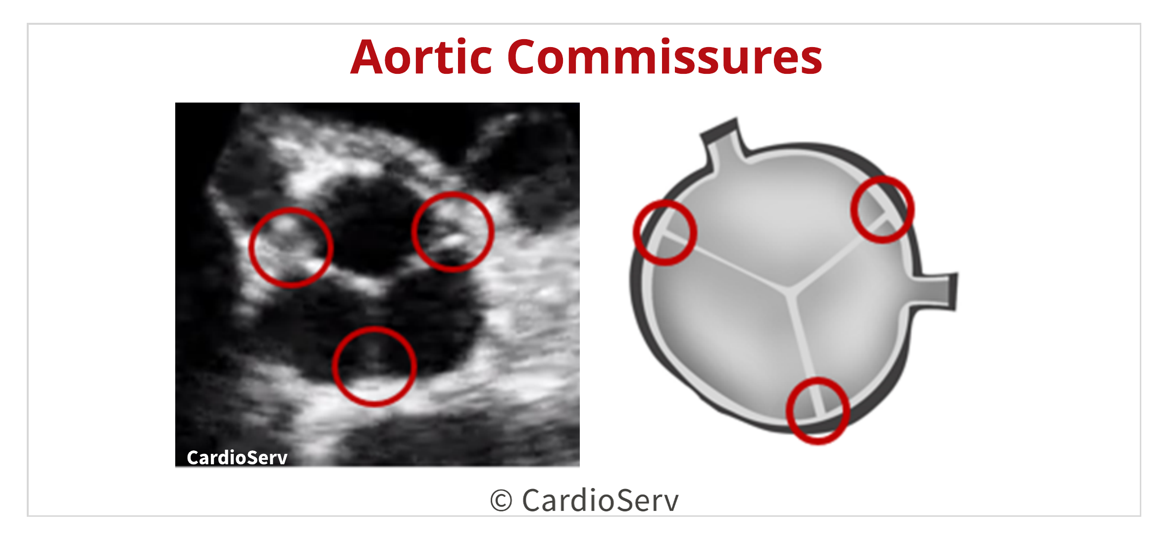
6,7 To date, larger studies that investigate the electrophysiological. The echo probe is placed within the esophagus and ultrasound is projected at the neighboring structures, such as the heart and aorta, which are in close proximity. The aortic valve is composed of 3 commissures.
🔴AORTIC STENOSIS 🔴 🔵 INTRODUCTION * The normal aortic valve consists of 3 symmetric cusps ️ Right Coronary Cusp (RCC), Left Coronary cusp (LCC) & Non- Coronary Cusp (NCC). Echocardiography is a dynamic assessment and it is important to examine structures through the entire cardiac cycle. The aortic valve normally has three cusps however there is some discrepancy in their naming.
The bottom, left video shows the direct visualization of the aortic valve, as viewed from the aorta, throughout the cardiac cycle. Start studying Echo Images. Given the unobstructed origin and benign course, no intervention was recommended.
‘non coronary cusp’. 6, 7 In the last decade, a few reports were published on focal AT originating from the non-coronary cusp (NCC). Three subtypes of cusp prolapse may be identified on echo:.
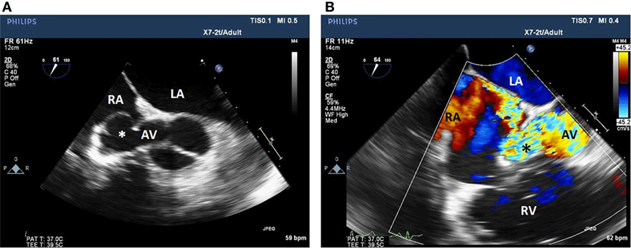
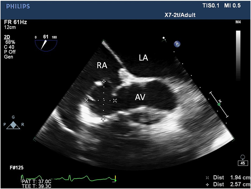
Although an LAA thrombus was absent, an incidental finding of a 5.3‐cm right coronary cusp (RCC) aneurysm was noted. Arrows depict direction of blood flow through the defect between the aorta and right atrium. Ninety to 95% of these congenital aneurysms originates in the right or non-coronary sinus and project into the right ventricle or into the right atrium.
During the operation, we observed a 30 × mm ruptured aneurysm that arose from the non. Intraoperative transoesophageal echocardiography showed that the commissure between the right coronary and non-coronary cusp of the aortic valve was detached from the aortic wall, with resultant right coronary cusp prolapse and severe aortic regurgitation. A bilateral tear in the right coronary and non-coronary cusp was found.
Left Ventricle wall segments. Right coronary cusp LCC:. Download high-res image (1MB) Download :.
Focal atrial tachycardias (ATs) have a predilection for specific areas within the atrium, such as the crista terminalis, 1 pulmonary vein ostia, 2 near the tricuspid and mitral annulus, 3, 4 the ostium of the coronary sinus (CS), 5 and para-Hisian regions. Aneurysm arising in the non-coronary sinus almost all rupture into the right atrium, and those arising in the right coronary sinus generally communicate with the right ventricle and occasionally with the right atrium. The mitral valve is also called the bicuspid valve and the left atrioventricular valve.
Atrial tachycardias (ATs) surrounding the region of the aortic coronary cusps have been reported in limited numbers of patients. AV SAX showing mobile mass attached to the non-coronary cusp Video 3:. Question 2 Which myocardial segment is denoted by the arrow?.
As the name bicuspid valve may suggest, the mitral valve is considered to have two primary leaflets:. (A) Biplane transesophageal echocardiography images (left panel, long-axis image;. The Raman spectra of the non-coronary cusp (NCC) were divided into two groups:.
Aortic valves typically have 3 cusps (right, left, and non-coronary), however, unicuspid, bicuspid, and quadricuspid valves can occur. Note again the non coronary cusp. Right posterior PV cusp 7.
The VSD shunt was restricted by the prolapsed cusp. 3b Ruptured non-coronary Sinus of Valsalva aneurysm. Through multimodality imaging, we present a young female with an anomalous RCA arising from the non-coronary cusp.
NCC = noncoronary cusp;. The commissures are the coaptation lines that run parallel between the leaflets. AV Short Axis – 3D view of the aortic valve from the ascending aorta showing mobile mass attached to the non-coronary cusp Video 4:.
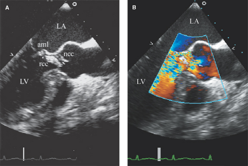
Some sources also advocate they be named as a left, right and posterior cusp. MV = mitral valve. Long-axis view — In the long axis view, typically at 110 to 140˚ from transverse (0˚) , the right and non-coronary cusps are visualized and the presence of any vegetations or leaflet prolapse is usually evident and regurgitant jet width can be ascertained.
945- and 970-cm −1 bands (traces 1,2) and 959- and 1,070-cm −1 bands (traces 3–5;. The interleaflet triangle formed between the right and non-coronary cusp is part of the “central fibrous body,” which represents the convergence of the two atrioventricular valves with the aortic valve. LCC =left coronary cusp;.
In the parasternal long-axis view the anterior mitral valve leaflet appears _____ and _____ then the posterior mitral leaflet. Crossref Medline Google Scholar;. Echocardiography found severe aortic regurgitation and aortic valve aneurysm of the non-coronary cusp going in and out of the left ventricular chamber.
After induction of anesthesia, an inflow duct for cardiopulmonary bypass (CPB) was placed in his right subclavian artery. Right Coronary Cusp (RCC) Noncoronary Cusp (NCC) Ascending Aorta (Size) Aortic Root (Diameter) Sinotubular Junction (Size) Right Pulmonary Artery (RPA) Left Atrium (Diameter) Coronary Sinus (Size). The patient was recommended cardioversion and underwent transesophageal echocardiography prior to the procedure to evaluate for left atrial appendage (LAA) thrombus.
15 Corrado D, Thiene G, Cocco P, Frescura C. OTHER SETS BY THIS CREATOR. Non-Coronary Sinus bovine pericardial patch (white arrow).
Cusp flail, whole cusp and partial cusp prolapse. 3c Excision of fistula and aneurysm with pericardial patching of the non-coronary sinus. Learn vocabulary, terms, and more with flashcards, games, and other study tools.
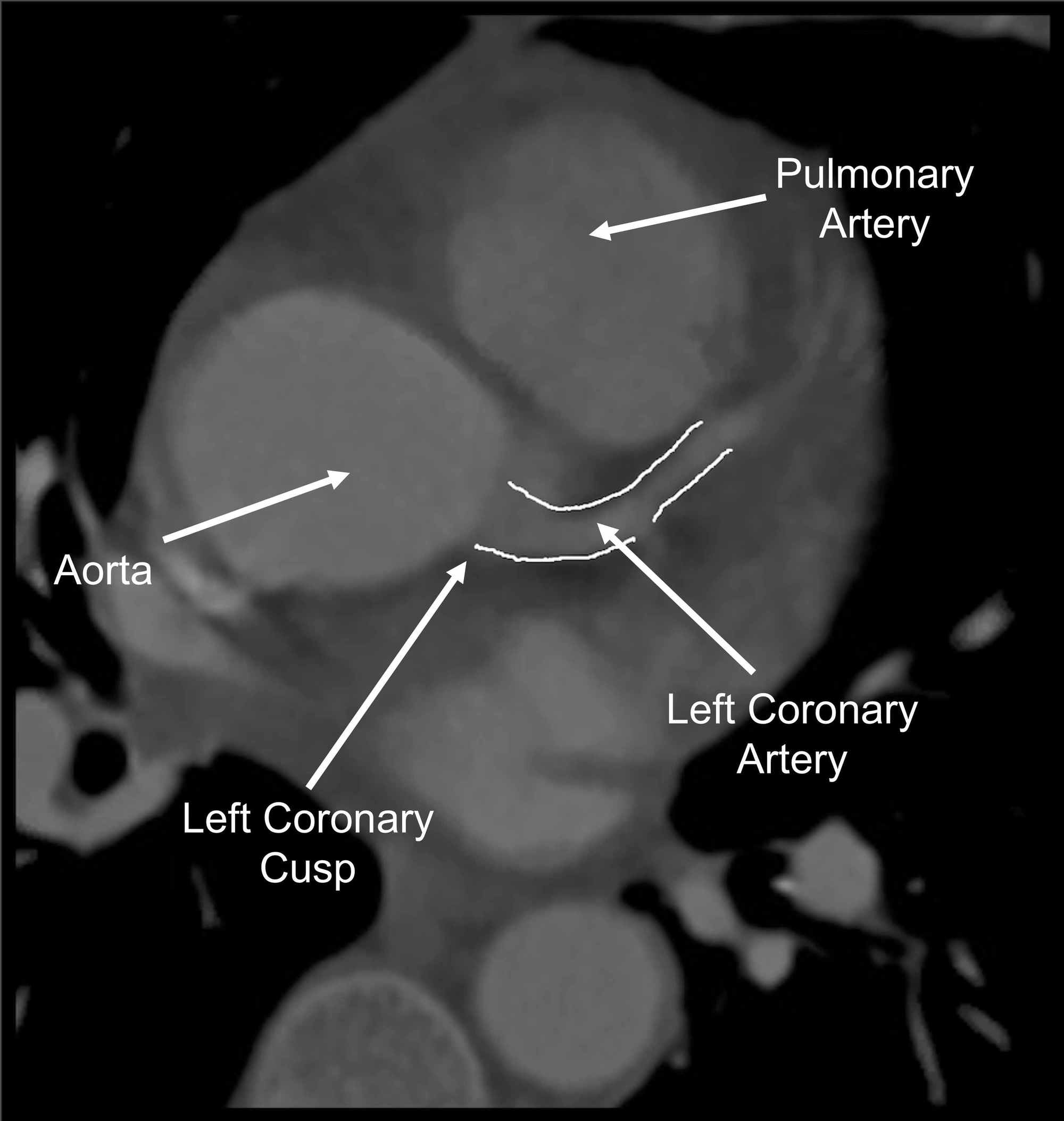
Left main coronary artery originating from the right sinus of Valsalva and coursing between the aorta and pulmonary trunk. P A S S Adult Echocardiography Workbook Ch 1 Anatomy and Physiology. Echocardiography is the initial modality employed in the assessment of proximal coronary arteries in children (Video 1).
Non coronary cusp To receive notifications about the YouTube videos and playlists from echocardiography, click the Subscribe button to. Non-coronary cusp aortic valve. Right panel, short-axis image) of prosthetic aortic valve at 2nd aortic valve replacement.
RCC, right coronary cusp. Quadricuspid aortic valves have 4 cusps where a single original cusp has split or failed to fuse in utero. Therefore, the patient underwent aortic valve replacement.
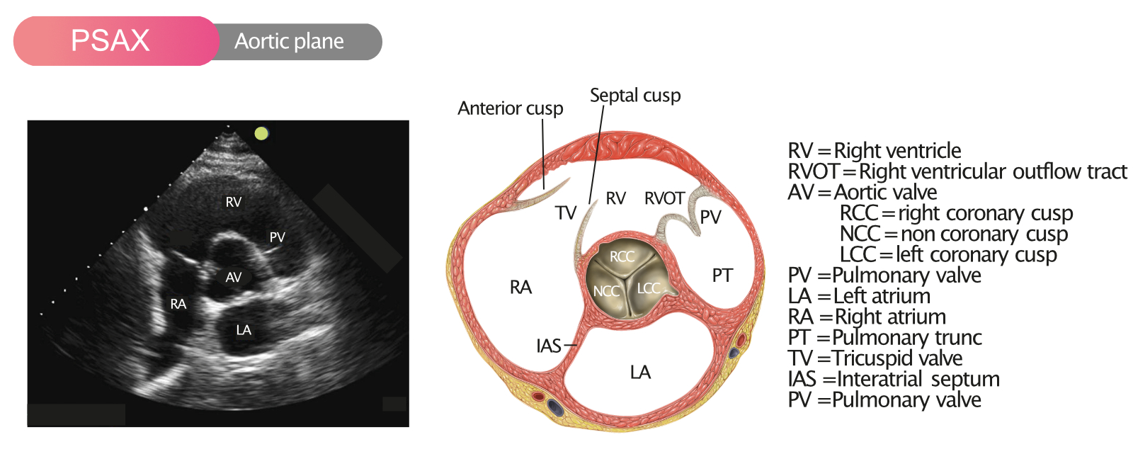
For the echocardiographer looking down at the aortic valve in the short axis view, the non-coronary cusp will be in the 7 o’clock position, the right coronary cusp will be in the 11 o’clock position and the left coronary cusp will be in the 3 o’clock position. 1 However, echo images may be sub-optimal in older patients with high body mass indices. The final cusp is named the non-coronary cusp and is positioned posteriorly relative to the other two cusps.
Transesophageal echocardiography improves image quality in many ways over transthoracic echo but at the expense of being an invasive procedure. 2.03 cm, aortic sinus:. The right coronary cusp, the left coronary cusp, and the non-coronary cusp.
AOV non coronary cusp 6. The aortic valve controls the flow of blood leaving the left ventricle and has three cusps:. Right coronary cusp aortic.
The interleaflet triangle between the non- and left coronary cusp is continuous with the anterior leaflet of the mitral valve. Figure 2C), suggesting that the calcification process in the NCC predominantly involved the formation of B-type carbonated HA (major) and β-tricalcium phosphate (minor). The aortic valve can be seen in this plane as two of the three aortic leaflets (typically the non coronary cusp and right coronary cusp) mark diastole (closed) and systole (open).
AV LAX showing mobile mass attached to the left or non-coronary cusp Video 2:. There was no evidence of aortic stenosis or regurgitation. 1 – 7 The location of successful ablation targets includes the noncoronary cusp (NCC) in most case series 1 – 5 and the left coronary cusp (LCC) in a few case reports.
The aortic valve was removed and replaced with a number 27 Carpentier Edwards prosthesis. The humble echocardiography can identify the origin* of coronary arteries in most persons. Improved resolution and precise details of coronary anatomy may be obtained by computed tomography angiography (CTA) with virtual.
In the long axis, the ascending aorta should be viewed from the valve to the right. He had significant prolapse of the non-coronary cusp, dilation of the corresponding sinus of valsalva, and severe aortic regurgitation. A qrS pattern in leads V1-V3.
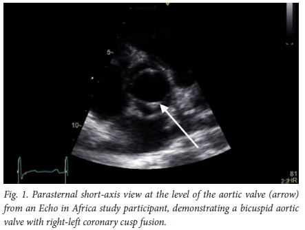
Cusp prolapse is diagnosed when the free edge of one or more aortic cusps overrides the plane of the aortic annulus. J Am Coll Cardiol. Echocardiography showed bicuspid aortic valve with fusion of the right and non-coronary cusp, unruptured aneurysm of left coronary sinus of Valsalva bulging into left atrium, gross dilation of aortic root (aortic annulus:.
While you’re answering the questions, take the time to appreciate the relative and absolute sizes of the cardiac structures, the global and regional function of the right and left ventricles, and the appearance of normal valves. The noncoronary cusp was involved most frequently in 56% (p < 0.01), followed by the right coronary cusp in 35%, and the left coronary cusp in 9%. AV Short Axis – 3D Cropped showing attachment point of the mobile mass at the base of the non.
Anesthetic Implications Of The Anomalous Origin Of Coronary Artery Aoca
Echocardiography Tutorial Aortic Valve
The Role Of Echocardiography In Transcatheter Aortic Valve Implantation Onishi Cardiovascular Diagnosis And Therapy
Www Jtcvs Org Article S0022 5223 10 4 Pdf

Echocardiography In General Practice 4 Views To Master Clinician S Brief
Sinus Of Valsalva To Right Atrial Fistula Heart

Korean Journal Of Anesthesiology

The Role Of Echocardiography In Aortic Valve Repair Vanoverschelde Annals Of Cardiothoracic Surgery

Aortic Valve Aneurysm Responsible For Acute Congestive Heart Failure And Histological Findings A Case Report Sciencedirect
Aortic Sclerosis Diagnosis Treatments Risk Factors

Echocardiography For The Surgeons Ppt Download

The Korean Journal Of Internal Medicine

A Tale Of Four Valves Outcome Of Brucella Endocarditis A Case Series Abstract Europe Pmc
Back To The Basics Aortic Valve Anatomy
Echo Assessment Of Aortic Valve Disease Dr Ferdous Assistant Registr

Guidelines For The Use Of Transesophageal Echocardiography To Assist With Surgical Decision Making In The Operating Room A Surgery Based Approach Journal Of The American Society Of Echocardiography

Parasternal Long Axis Views 2 Of 4

View Image

Figure6 Multimodality Imaging Of Thoracic Aortic Diseases In Adults Jacc Cardiovascular Imaging

Parasternal Short Axis Views 2 Of 5

Aortic Valve Stenosis A Practical Approach To Clinical Echocardiography 1st Edition
Echocardiographic Diagnosis Of Rare Pathological Patterns Of Sinus Of Valsalva Aneurysm

Rheumatic Aortic Valve Disease With Mitral Stenosis A Case Report

Aortic Anatomy

Parasternal Long Axis Views 2 Of 4

Jaypeedigital Ebook Reader

Standard Transthoracic Echocardiogram Complete Imaging Protocol Ecg Echo

Valsalva Sinus Perforation Following Valve Dislodgement During Transcatheter Aortic Valve Replacement Jacc Cardiovascular Interventions
Back To The Basics Aortic Valve Anatomy

Echocardiopro
Aortic Valve Echopedia
Frontiers Incidental Finding Of An Aorto Right Atrial Fistula In A Patient Undergoing Repair Of A Sinus Of Valsalva Aneurysm Medicine

Aortic Stenosis Cancer Therapy Advisor

Normal Valves Chapter 5 Core Topics In Transesophageal Echocardiography

Parasternal Short Axis Views 2 Of 5

Quadricuspid Aortic Valve Los Angeles Echo Society Facebook

Parasternal Short Axis Views 2 Of 5

Short Axis View With Labeling Of The Non Ncc Left Lcc And Right Download Scientific Diagram

Cardiac Transthoracic Echocardiography Tte Summary And Labeled Views Rk Md
Echocardiography Of Aortic Stenosis

Parasternal Long Axis Icu Echo

Valves Echocardiography Radiology Key

Noncoronary Sinus Of Valsalva Aneurysm Resembling A Cystic Cardiac Mass Sonsoz Echocardiography Wiley Online Library
Anomalous Origin Of Right Coronary Artery From The Non Coronary Cusp Demonstrated By Echocardiography And Ct Angiography Springerlink
Infective Endocarditis Anesthesia Key
Aortic Valve Stenosis Ecg Echo

Scan Views Echocardiografie

Preoperative Aortic Annulus Size Assessment By Transthoracic Echocardiography Compared To The Size Of Surgically Implanted Aortic Prostheses In Echo Research And Practice Volume 6 Issue 2 19

Transesophageal Echo
Echocardiographic Diagnosis Of Rare Pathological Patterns Of Sinus Of Valsalva Aneurysm

Echo Protocol Diagram Quizlet

Aortic Valve Stenosis A Practical Approach To Clinical Echocardiography 1st Edition
Transthoracic Echocardiography Tte Showing Focal Thickening On The Download Scientific Diagram
Q Tbn 3aand9gcr Avjgahg5avg40gipueafa2lk1otokdjpysk0kqxi1gkd1wpn Usqp Cau
Www Cvcasejournal Com Article S2468 6441 18 9 Pdf

Bicuspid Aortic Valve Intechopen
Aortic Valve Quiz Echocardiography Ultrasound And Perioperative Medicine

Systematic Echocardiographic Assessment Of Aortic Regurgitation What Should The Surgeon Know For Aortic Valve Repair Berrebi Annals Of Cardiothoracic Surgery

Non Coronary Cusp Dr S Venkatesan Md
Lessons Of The Month 3 Gone But Not Forgotten Osler A Reminder Of The Syndrome Not Bearing His Name Rcp Journals

View Image

Multiple Papillary Fibroelastomas Of The Aortic Valve Detected By Real Time Three Dimensional Transesophageal Echocardiographic Images Sciencedirect

Echocardiography And The General Physician Postgraduate Medical Journal
Valvular Heart Disease Radiology Key

Parasternal Short Axis Basal Cardiac Sonography Ultrasound Diagnostic Medical Sonography

The Role Of Echocardiography In Aortic Valve Repair Vanoverschelde Annals Of Cardiothoracic Surgery
Role Of Echocardiography In Transcatheter Aortic Valve Implantation Springerlink

Transesophageal Echo

Sinus Of Valsalva To Right Atrial Fistula Heart
1

Aortic Valve Short Axis View Ac Acoronary Lc Left Coronary Rc Download Scientific Diagram

Parasternal Short Axis Views Echocardiography

Epos Trade
Colour Doppler Echo Quiz Discussion All About Cardiovascular System And Disorders
Transesophageal Echocardiography Thoracic Key
Transesophageal Echocardiography Long Axis View Of Aorta Showing Download Scientific Diagram
Rupture Of Noncoronary Sinus Of Valsalva Into Right Atrium In The Fifth Decade Of Life Rajan R Amin O Soman B Dashti R Al Jarallah M Heart India

Figure 1 From Bicuspid Aortic Valve An Unusual Cause Of Aneurysm Of Left Coronary Sinus Of Valsalva Semantic Scholar

3d Transoesophageal Echocardiography In The Tavi Sizing Arena Should We Do It And How Do We Do It In Echo Research And Practice Volume 4 Issue 1 17
Jafib

Echocardiography Evaluation Of Aortic Regurgitation Echocardiography
Aortic Valve Repair
Congenital Bicuspid Aortic Valve Differential Prevalence Across Different South African Population Groups

The Role Of Echocardiography In Transcatheter Aortic Valve Implantation Onishi Cardiovascular Diagnosis And Therapy
3d Echo In Preoperative Assessment Of Aortic Cusps Effective Height

Multiple Ruptured Aneurysm Of Left Sinus Of Valsalva A Rare Entity Sarupria A Kapoor Pm Kiran U Hote M Ann Card Anaesth

Transesophageal Echocardiography Instrumentation And System Controls

Ultrasound Board Review

Scan Views Echocardiografie

Use Of Intracardiac Echocardiography In Interventional Cardiology Circulation

Ultrasound Board Review
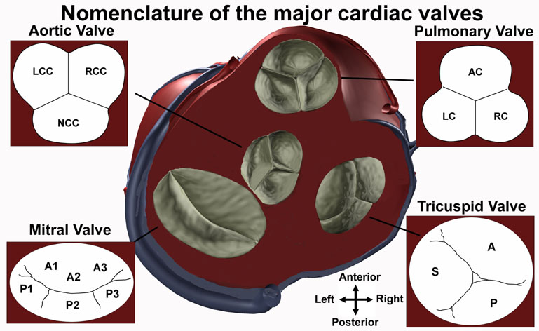
Anatomy Tutorial Cardiac Valve Nomenclature Atlas Of Human Cardiac Anatomy
Q Tbn 3aand9gcrx9u2kenrcm8nl6bvxsxxzxnmgk2q Vnkh5g0pkqqepdyknubf Usqp Cau
Two Dimensional Transesophageal Echocardiogram Image Of The Aortic Download Scientific Diagram

View Image

The Heart Of A Horse 3 D Echocardiographic Analysis Of The Equine Aortic Valve Inquiry Journal

Figure 1 From Aortic Regurgitation Echocardiographic Diagnosis Semantic Scholar
Cureus Incidentally Diagnosed Anomalous Right Coronary Artery With An Interarterial Course Presenting As Chest Pain


