Lv Non Compaction Echo Criteria
Q Tbn 3aand9gcrz9c0efozcuepf Mhbuq5q7hnpdepabyipbkxkwxe 93qi Cpu Usqp Cau
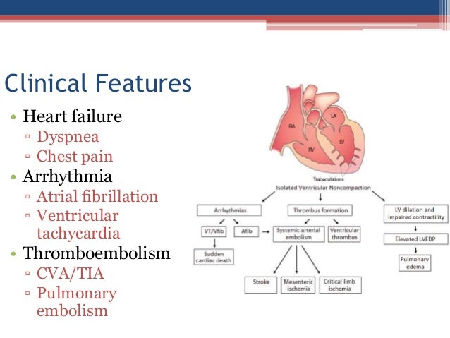
Noncompaction Cardiomyopathy

Isolated Left Ventricular Non Compaction Cardiomyopathy In Adults Sciencedirect

Myocardial Deformation Pattern In Left Ventricular Non Compaction Comparison With Dilated Cardiomyopathy Abstract Europe Pmc

Figure 4 42 From Left Ventricular Noncompaction Imaging Findings And Diagnostic Criteria Semantic Scholar

Improvement In Systolic Function In Left Ventricular Non Compaction Cardiomyopathy A Case Report Sciencedirect
Echocardiography showed L V non-compaction with.

Lv non compaction echo criteria. Patients who presented with VT had evidence of abnormal electroanatomic substrate involving the mid- to apical segments of the LV, which matched the noncompacted myocardial segments identified by preprocedural magnetic resonance imaging or echocardiography. Do we need more stringent criteria for the diagnosis of left ventricular non-compaction in athletes?. Left ventricular noncompaction 1.
Heart Br Card Soc. Perfusion of the intertrabecular spaces from the ventricular cavity is present at end-diastole on colour Doppler echocardiography or contrast echocardiography Refinement of echocardiographic criteria for left ventricular noncompaction Claudia Stöllberger,Birgit Gerecke,Josef Finsterer,Rolf Engberding.International Journal of Cardiology 165. It can be associated with left ventricular dilation or hypertrophy, systolic or diastolic dysfunction, or both, or various forms of congenital heart disease.
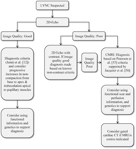
Many imaging modalities have been evaluated for the diagnosis of left ventricular noncompaction (LVNC), including echocardiography, 1 angiography, 2 computed tomography,3, 4 and magnetic resonance imaging.4, 5 However, echocardiography complemented with color Doppler flow imaging is currently the diagnostic modality of choice. Current echocardiographic criteria for diagnosis typically include:. Eight women showed sufficient trabeculations to fulfill criteria for LV noncompaction.
1 We present a series of adult patients with suspected LVNC on. In echocardiography, the fetal LV myocardium is a two-layered structure:. Left ventricular (LV) hypertrabeculation is defined by the presence of three or more trabeculations apically and up to the level of papillary muscles, seen in one echocardiographic view.1 It can be distinguished from left ventricular non-compaction (LVNC) by the absence of thin compacted myocardial layer.
Gati S(1), Rajani R(2), Carr-White GS(2), Chambers JB(2). Also called insulated non compaction of the ventricular myocardium (INVM), it is a rare form of congenital heart disease in which the tissue of the ventricular myocardium is not well constructed in terms of texture. Left ventricular noncompaction is a rare unclassified cardiomyopathy with markedly prominent apical trabeculae with deep intertrabecular recesses (Fig.
42 are generally accepted (Table 3). An uninformative test has a sensitivity and specificity of 50%. Due in part to improved imaging with echocardiography and magnetic resonance imaging, clinical awareness and appreciation for the marked heterogeneity of this disorder is increasing.
The objectives of this article are to review the imaging findings of left ventricular noncompaction (LVNC) at echocardiography, cardiac MRI, and MDCT;. Furthermore, the criteria are indirect, assessing morphologicalabnormalities.Aftercarefulevaluation of all criteria, the most important echocardiographic criterion remains a noncompacted/compacted ratio >2.0 in end-systole(49,50). Left ventricular noncompaction (LVNC) cardiomyopathy is characterized by prominent myocardial trabeculations and deep recesses.
This causes channels to form in the heart muscle, called trabeculations. The noncompacted layer is thinner than the compacted layer in the anterior wall, but thicker than the compacted layers in the posterior, lateral, and inferior wall. Left Ventricular Non-Compaction Case Studies Matt Umland, ACS, RDCS, FASE Aurora Health Care Milwaukee, WI Left Ventricular Noncompaction Cardiomyopathy • 1926 Grant - Malformed heart of a child • 1975 Dusek - Spongy Myocardium • 1984 Englberding – Echo Diagnosis of Myocardial Sinusoids • 1986 Jenni – Biventricular Sinusoids.
And to describe pitfalls that can lead to misinterpretation of findings of LVNC. Isr Med Assoc J 09;11:313-4. Reappraisal of current diagnostic imaging modalities.
At least some of the inconsistency is due to the fact that non-compaction of the left ventricle is mostly diagnosed by morphological criteria on non-invasive imaging (e.g. Presence of multiple echocardiographic trabeculations, multiple deep intertrabecular recesses communicating with the ventricular cavity, a 2-layered structure of the endomyocardium with an increased noncompacted to compacted ratio. Noncompacted myocardium should be measured in a plane perpendicular to the compacted myocardium.
Echocardiography and cardiac MRI), and no universally accepted criteria exist. During the postpartum follow-up period of 24±3 months. Non-compaction is diagnosed when the trabeculations are more than twice the thickness of the underlying ventricular wall.
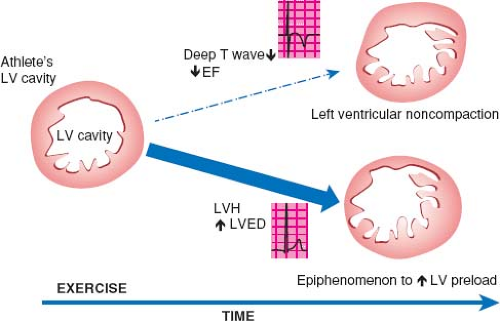
Increased left ventricular trabeculation in highly trained athletes:. Left ventricular noncompaction (LVNC) is a distinct phenotype characterized by prominent LV trabeculae and deep intertrabecular recesses .LVNC was previously also called spongy myocardium or hypertrabeculation syndrome but these terms should not be used interchangeably with LVNC .This review will focus on clinical manifestations and diagnosis of LVNC as an isolated disorder. What is left ventricular non-compaction (LVNC)?.
Left ventricular non-compaction, the most recently classified form of cardiomyopathy, is characterised by abnormal trabeculations in the left ventricle, most frequently at the apex. Our echocardiography laboratory was consulted to determine whether a patient's echocardiogram would fulfill the criteria of left ventricular noncompaction cardiomyopathy (LVNC). All 38 patients had a diagnosis of LV non-compaction on cardiac MRI, using criteria of non-compacted/compacted myocardium > 2.5 to 1.0 and deep LV trabeculations.
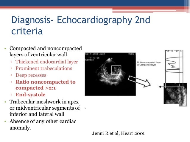
During development, the heart muscle is a sponge-like network of muscle fibers. 3 The clinical diagnosis is predominantly reliant on three proposed echocardiographic criteria and based on an increased ratio of the noncompacted inner. Stöllberger et al base their diagnosis of left ventricular noncompaction on Jenni et al's echocardiographic diagnostic criteria 3:.
Whereas echocardiography criteria is the frequently used for diagnosis, the three criteria are non-specific and overlapping. Left ventricular non compaction (LVNC) is a type of cardiomyopathy which is characterized by the presence of prominent trabeculations in the left ventricle with deep recesses between the trabeculations and a thin compacted myocardial layer. Sports cardiology institutions in the UK and France.
Thomas' NHS Foundation Trust, London, United Kingdom. To investigate the prevalence and significance of increased left ventricular (LV) trabeculation in highly trained athletes. LV solid body rotation with near absent LV twist and LV geometry may be new quantitative functional diagnostic criteria for the diagnosis of LVNC .24, 25, 26 Three-dimensional imaging in selected cases has allowed better characterization of noncompacted and compacted myocardium (Figure 9, Video 7).
These pieces of muscles are called trabeculations. These are best visualized on color flow Doppler of the left ventricle using apical windows. Comparison of Echocardiographic Diagnostic Criteria of Left Ventricular Noncompaction in a Pediatric Population.
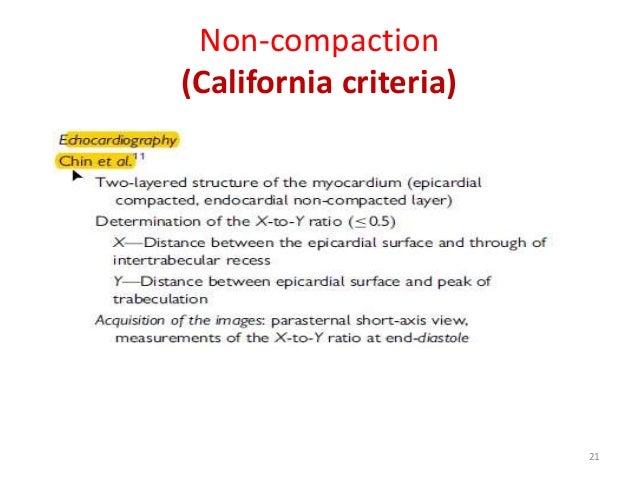
In left ventricular non-compaction cardiomyopathy (LVNC) the lower left chamber of the heart, called the left ventricle, contains bundles or pieces of muscle that extend into the chamber. Mutations in Cypher/ZASP in patients with dilated cardiomyopathy and left ventricular non-compaction. A) >3 trabeculae projecting from the left ventricular wall distal to the papillary muscles, visible in a single echocardiographic plane;.
NCCM is characterized by excessive trabeculations typically involving the left ventricle (LV) with >2:1 ratio of noncompacted:compacted myocardium. Advances in cardiovascular imaging and widespread availability of imaging technology have led to an increase in the diagnosis of LVNC imposing a need for evidence-based imaging diagnostic criteria. Vatta M, Mohapatra B, Jimenez S, et al.
More recently, Chin et al 2 reported the isolated form. 10.1136/heartjnl-12- Crossref Medline Google Scholar. Left ventricular noncompaction is a rare cardiomyopathy that should always be considered as a possible diagnosis because of its potential complications.
LVNC is a condition of the heart where the walls of the left ventricle (the bottom chamber of the left side of the heart) are non-compacted. These criteria include a bilayered myocardium, a noncompacted to compacted ratio >2 :. Presenting with Left Ventricular Non Compaction (LVNC, Group 1) or Idiopathic Dilated Cardiomyopathy (DCM, Group 2) At distance of an acute heart failure thrust (> 1 month) Newly diagnosed (less than 6 months) Diagnosis confirmed by echocardiography associated or not with a Magnetic Resonance Imaging (MRI) confirmed after central review.
(1)Department of Cardiology and Cardiac Imaging, Guy's and St. In very low pre-test probabilities consistent with the reported left ventricular noncompaction (LVNC) prevalence of 0.014% to 0.5%, neither cardiac magnetic resonance (CMR) criteria are very informative. To discuss diagnostic criteria for and the advantages and limitations of these imaging techniques;.
1-3 The clinical spectrum of the disorder ranges from being completely asymptomatic to progressive left ventricular (LV) systolic impairment, a tendency to fatal arrhythmias and systemic thromboembolic events. 1146 athletes aged 14-35 years (63.3% male), participating in 27 sporting disciplines, and 415 healthy controls of similar age. The diagnosis of left ventricular (LV) non-compaction has profound implications for patients, yet no uniform criteria for its diagnosis by echocardiography exist.
Cross sectional echocardiographic study. Left Ventricular Non Compaction Cardiomyopathy (LVNC) Raysa Morales-Demori, MD. Left ventricular noncompaction 2.
2 By definition, noncompaction pertains to the LV but may also involve the right ventricle, as either a. None of the 22 patients with both 2-D echo and cardiac MRI exams had a diagnosis of LV non-compaction on their 2-D echo study. Tissue Doppler imaging studies of regional deformation seem to distinguish isolated left ventricular noncompaction (iLVNC) from DCM , and 2-dimensional speckle-tracking echocardiography seems to detect myocardial dysfunction in patients with LVNC and normal LV function by using conventional methods.
MedlinePlus - Health Information from the National Library. Non-compaction cardiomyopathy (NCM) is a myocardial disorder, which is thought to occur due to the failure of left ventricle (LV) compaction during embryogenesis, leading to distinct morphological characteristics in the ventricular chamber.1 It was first described about 80 years ago, in association with complex congenital heart diseases. A total of 8 patients (%) had left ventricular (LV) systolic dysfunction (mean ejection fraction 40% ± 13%).
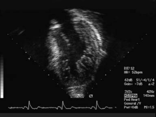
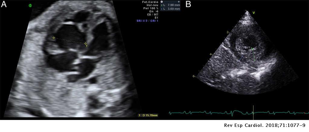
In normal human hearts of children and adults the left ventricle (LV) has up to 3 prominent trabeculations and is, thus, less trabeculated than the right ventricle 1, 2.Rarely, more than 3 prominent trabeculations that is the so-called LV noncompaction of ventricular myocardium (NVM) can be found at autopsy and by various imaging techniques including echocardiography and MRI etc. The MRI diagnostic criteria proposed by Petersen et al. Images of the left ventricle showed a 2-layer structure with a compacted, thin epicardial band and a much thicker noncompacted endocardial layer of trabecular meshwork.
Left ventricular noncompaction (LVNC) is a distinct phenotype characterized by prominent LV trabeculae and deep intertrabecular recesses .LVNC was previously also called spongy myocardium or hypertrabeculation syndrome but these terms should not be used interchangeably with LVNC .This review will focus on management of LVNC as an isolated disorder distinct from other clinical. Left ventricular noncompaction (LVNC) was first recognized in 1932 1 but was not officially described until 1990. Adult left ventricular noncompaction:.
Jenni criteria (Heart 07). We sought to find additive tools comparing the longitudinal strain characteristics …. Left ventricular non-compaction, also known as LVNC, spongy myocardium or hypertrabeculation syndrome, is a pathologic cardiac condition in which the myocytes exhibit a “spongy” appearance.
Diagnosis can be made by echocardiography;. Left ventricular non-compaction, cardiomyopathy, trabeculated left ventricular (LV), non-compacted endocardial layer, diagnosis, clinical management. Left ventricular noncompaction (LVNC) cardiomyopathy is morphologically characterized by prominent myocardial trabeculations and deep recesses.
2 Thought to be secondary to the arrest of normal myocardial development, LVNC results in multiple deep trabeculations in the left ventricle. 4 Left ventricular. Epub 17 Aug 3.
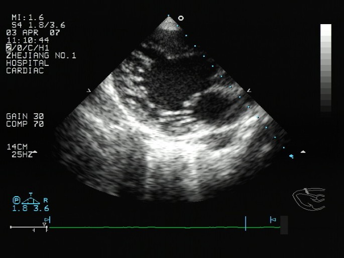
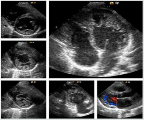
Two-dimensional apical four chamber and parasternal short axis images at the level of the ventricles show dilatation of both ventricles, multiple trabeculae and intertrabecular recesses in inferior, lateral, anterior walls, middle and apical portions of the septum and apex of the left ventricle. The precise stage of development and the natural history of the disorder are not fully understood. LVNC describes a ventricular wall anatomy characterized by the presence of disproportionate, prominent left ventricular (LV) trabeculae, a thin compacted layer, and deep intertrabecular recesses that are in continuity with the LV cavity and separated from the epicardial coronary arteries.
This gives the left ventricle a characteristic 'spongy' look (a bit like honeycomb). Left ventricular non-compaction (LV NC) is characterized by abnormal trabeculations that are mainly at the LV apex. 3 Clinical manifestations vary widely from asymptomatic to progressive deterioration resulting in heart failure.
The value of cardiac magnetic resonance imaging in the diagnosis of isolated non-compaction of the left ventricle. Hypertrabeculation is observed more often in competitive athletes, specifically in Afro. An end-diastolic ratio between noncompacted and compacted layers greater than 2.3 is considered diagnostic of myocardial noncompaction.
Affected individuals are at risk of left or right. Distinction between LV NC and non-specific dilated cardiomyopathies (DCMs) remains often challenging. The search for deformation indexes or early LV dysfunction in NC hearts is the expression of the clinical need to go beyond morphology in establishing whether the NC anatomy coincides with a.
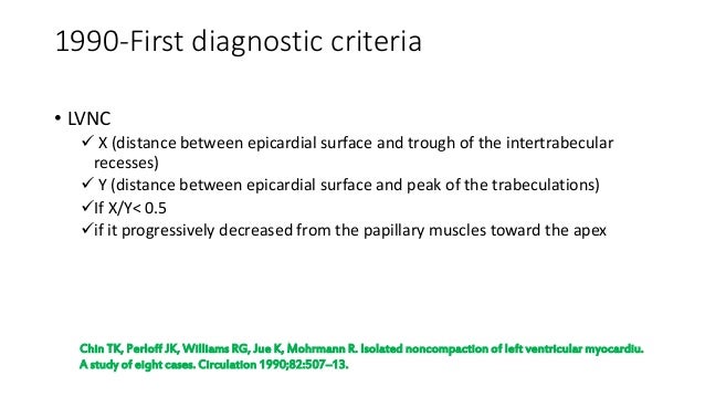
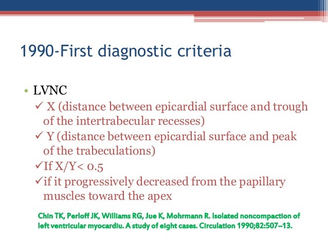
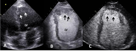
1, communication with the intertrabecular space demonstrated by Doppler, absence of coexisting cardiac abnormalities, and presence of multiple prominent trabeculations in end-systole 22. 1990-First diagnostic criteria • LVNC X (distance between epicardial surface and trough of the intertrabecular recesses) Y (distance between epicardial surface and peak of the trabeculations) If X/Y< 0.5 if it progressively. The endocardial noncompact myocardium (NC) with higher echo and the epicardium compact myocardium (C) with lower echo.
Through ECHO window 3. Isolated left ventricular non-compaction (LVNC) is a morphological abnormality of excessive trabeculation of the LV, often complicated by ventricular dysfunction, arrhythmias and cardioembolism. B) intertrabecular spaces perfused from the ventricular cavity (visualized by.

Adult Left Ventricular Noncompaction Jacc Cardiovascular Imaging

Masking And Unmasking Of Isolated Noncompaction Of The Left Ventricle With Real Time Contrast Echocardiography Circulation Cardiovascular Imaging

Catastrophic Stroke In A Patient With Left Ventricular Non Compaction In Echo Research And Practice Volume 5 Issue 3 18
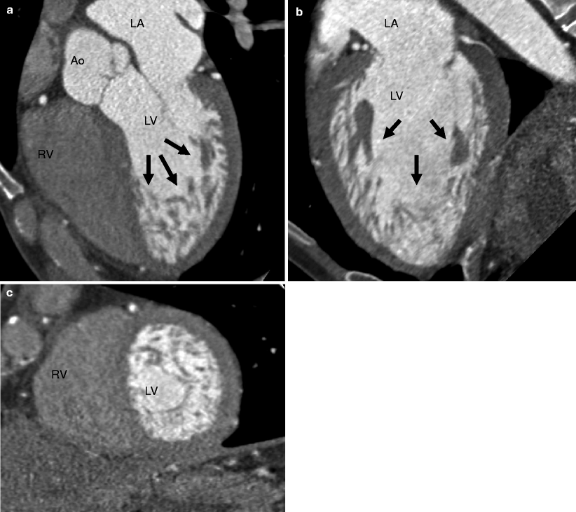
Left Ventricular Noncompaction Lvnc Radiology Key

Noncompaction Cardiomyopathy Case Presentation With Cardiac Magnetic Resonance Imaging Findings And Literature Review Saeedan Mb Fathala Al Mohammed Tlh Heart Views

Proposed Diagnostics Criteria Of Left Ventricular Non Compaction Download Table

Isolated Left Ventricular Noncompaction Cardiomyopathy A Transient Disease
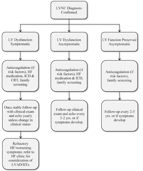
Left Ventricular Non Compaction A Review Of Literature On Clinical Status And Meta Analysis Of Diagnostic And Clinical Management Methods

Pedi Cardiology Lv Non Compaction
Www Internationaljournalofcardiology Com Article S0167 5273 09 4 Pdf
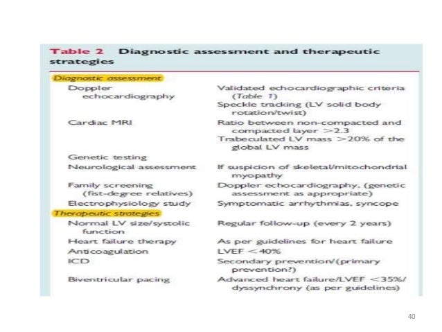
Left Ventricular Non Compaction

Echocardiographic Comparison Between Left Ventricular Non Compaction And Hypertrophic Cardiomyopathy Sciencedirect

Wall Thickness Measurements In Lv Non Compaction Para Sternal Short Download Scientific Diagram

Isolated Left Ventricular Noncompaction In Sub Saharan Africa A Clinical And Echocardiographic Perspective Circulation Cardiovascular Imaging
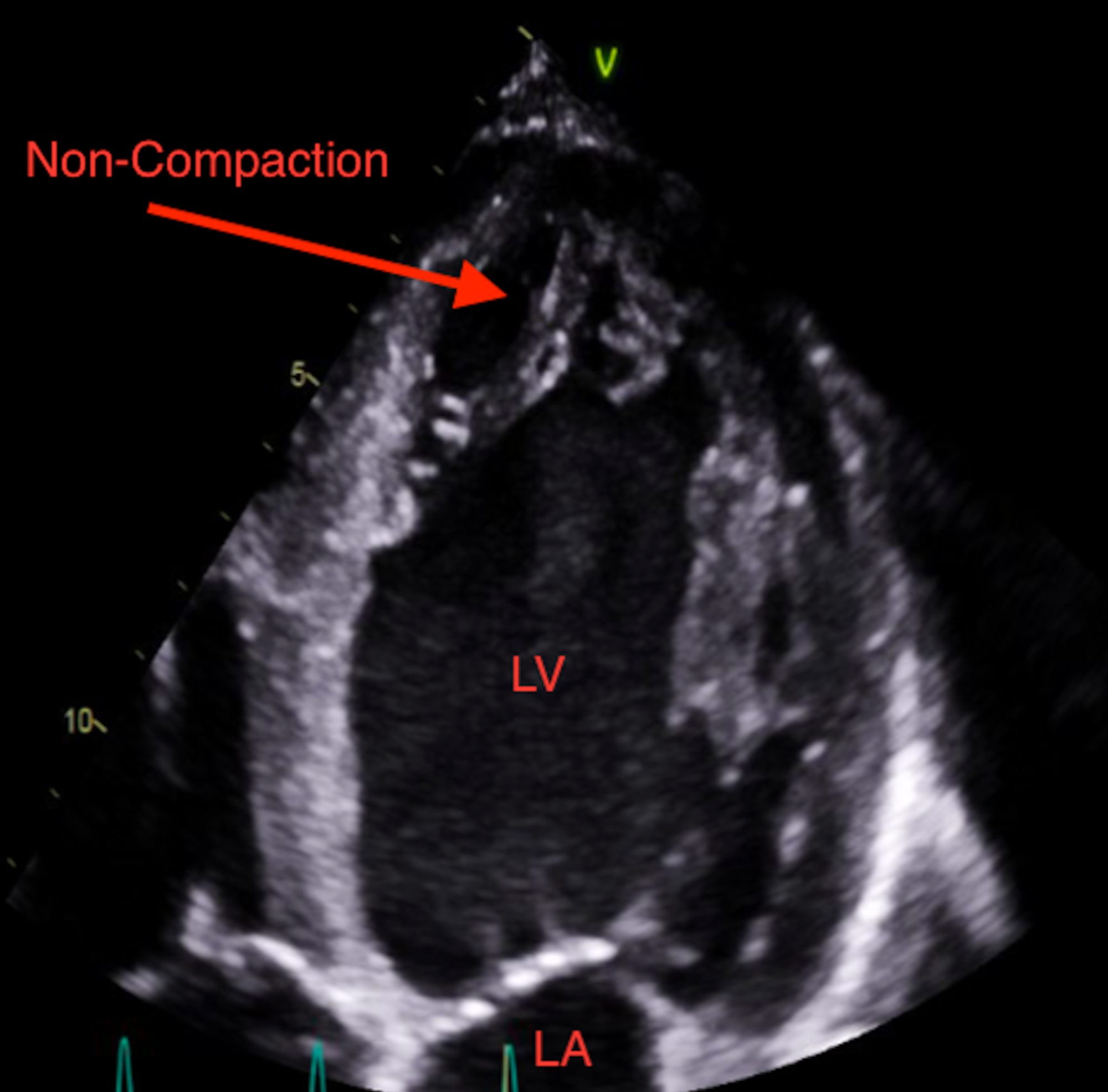
Cureus Heart Failure Secondary To Left Ventricular Non Compaction Cardiomyopathy In A 26 Year Old Male

Clinical And Echocardiography Features Of Diagnosed In Adulthood Isolated Left Ventricular Noncompaction A Case Series Study Huang Wh Sung Kt Tsai Jp Lo Ci Hsiao Cc Kuo Jy Su Ch Chen Mr
Q Tbn 3aand9gctgvbjhjzmvzgqtpdqugi0pfi2jttwcg9oim77pctd0oyxhq5ib Usqp Cau

Echocardiographic Criteria Of Lv Non Compaction Chin Criteria 1990 Absence Of Any Other

Assessment Of Left Ventricular Non Compaction In Adults Side By Side Comparison Of Cardiac Magnetic Resonance Imaging With Echocardiography Sciencedirect

British Cardiovascular Society

Non Compaction Of The Left Ventricle Radiology Reference Article Radiopaedia Org
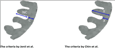
Left Ventricular Non Compaction A Review Of Literature On Clinical Status And Meta Analysis Of Diagnostic And Clinical Management Methods

Isolated Left Ventricular Noncompaction In Sub Saharan Africa A Clinical And Echocardiographic Perspective Circulation Cardiovascular Imaging

Table 1 From Diagnosis Of Left Ventricular Non Compaction In Patients With Left Ventricular Systolic Dysfunction Time For A Reappraisal Of Diagnostic Criteria Semantic Scholar

Echocardiogram Lv Non Compaction Youtube

Left Ventricular Noncompaction Cardiomyopathy Shemisa Cardiovascular Diagnosis And Therapy
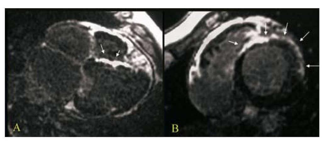
Late Gadolinium Enhancement In Non Compaction Cardiomyopathy Revista Espanola De Cardiologia English Edition

Reproducibility Of Echocardiographic Diagnosis Of Left Ventricular Noncompaction Thoracic Key

Isolated Left Ventricular Noncompaction Cardiomyopathy A Transient Disease
Www Asecho Org Wp Content Uploads 18 03 Umland Case Studies Left Ventricular Noncompaction Pdf
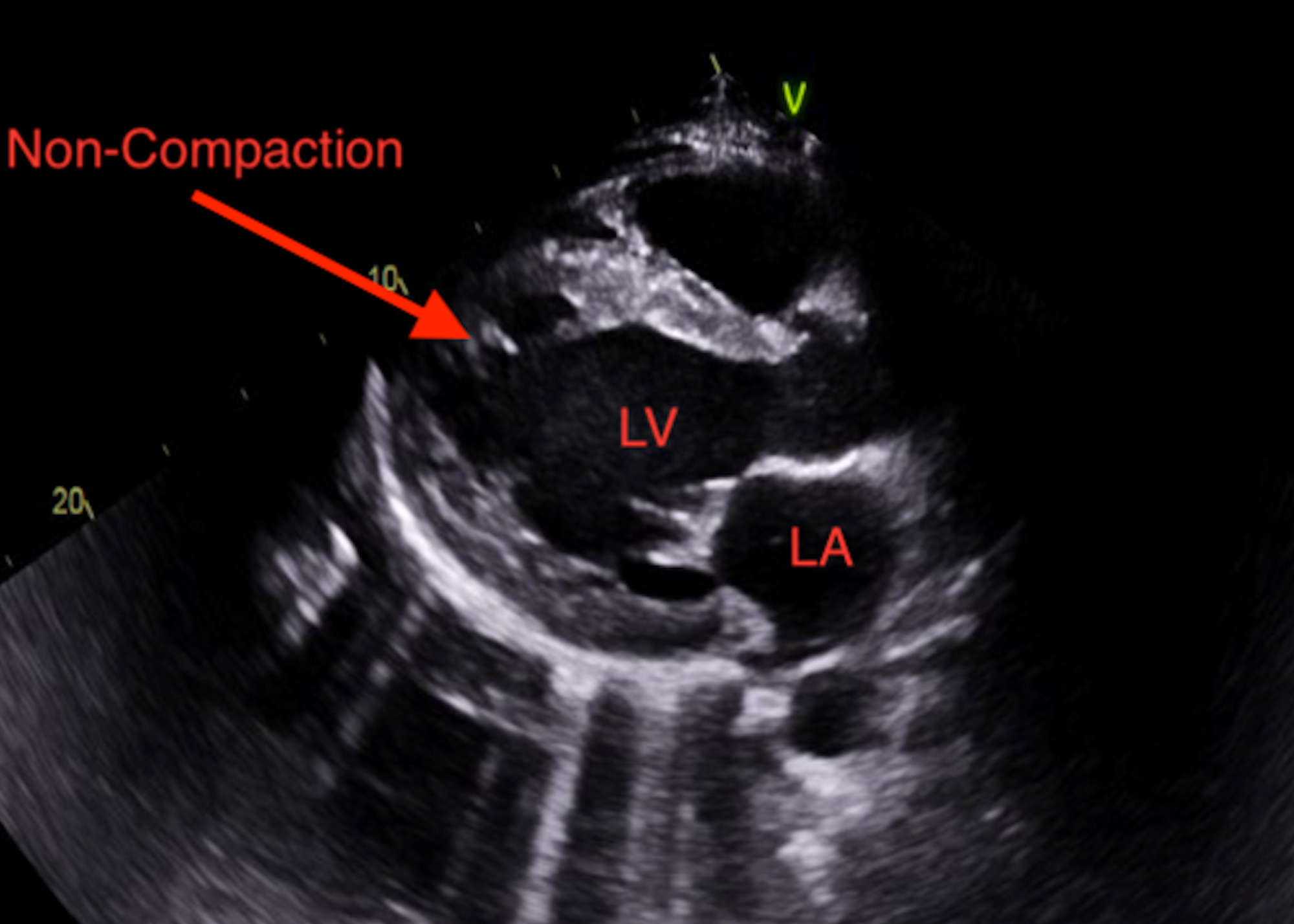
Cureus Heart Failure Secondary To Left Ventricular Non Compaction Cardiomyopathy In A 26 Year Old Male

Left Ventricular Trabeculations In Athletes American College Of Cardiology

Clinical And Echocardiography Features Of Diagnosed In Adulthood Isolated Left Ventricular Noncompaction A Case Series Study Huang Wh Sung Kt Tsai Jp Lo Ci Hsiao Cc Kuo Jy Su Ch Chen Mr

Left Ventricular Trabeculations In Athletes American College Of Cardiology

Left Ventricular Non Compaction Cardiomyopathy The Lancet

Left Ventricular Non Compaction Cardiomyopathy Lvnc

Isolated Ventricular Noncompaction Cardiomyopathy Presenting As Recurrent Syncope

Proposed Diagnostics Criteria Of Left Ventricular Non Compaction Download Table
Left Ventricular Non Compaction A Review Of Literature On Clinical Status And Meta Analysis Of Diagnostic And Clinical Management Methods

Reversible De Novo Left Ventricular Trabeculations In Pregnant Women Circulation

Echocardiography Findings In Common Primary And Secondary Cardiomyopathies Intechopen
Www Sads Org Sads Media International Conference Items 17 slides Sads Lvnc 17 Aj Pdf
Left Ventricular Noncompaction

Limitations In The Diagnosis Of Noncompaction Cardiomyopathy By Echocardiography
Www Asecho Org Wp Content Uploads 18 03 Umland Case Studies Left Ventricular Noncompaction Pdf
Echocardiography In The Diagnosis Left Ventricular Noncompaction Cardiovascular Ultrasound Full Text
A Patient With Abnormalities Of The Coronary Arteries And Non Compaction Of The Left Ventricular Myocardium Resulting In Ischaemic Heart Disease Symptoms Dabek Folia Morphologica

Left Ventricular Noncompaction A Distinct Genetic Cardiomyopathy Sciencedirect
Left Ventricular Non Compaction

Limitations In The Diagnosis Of Noncompaction Cardiomyopathy By Echocardiography
Q Tbn 3aand9gcqov7ennwh8lrzv70nehvwqjhj4ynpggm Oqkrwudsaj1hxqcy5 Usqp Cau

Left Ventricular Noncompaction Cardiomyopathy Shemisa Cardiovascular Diagnosis And Therapy

Multiple Thrombi In The Lv In Non Compaction Cardiomyopathy American College Of Cardiology

Non Compaction Cardiomyopathy Heart
Left Ventricular Noncompaction Cardiomyopathy Thoracic Key

Contrast And 3d Echocardiography In Lv Non Compaction Apical Four Download Scientific Diagram

Color Doppler In Lv Non Compaction Apical Four Chamber Views With Download Scientific Diagram
Left Ventricular Noncompaction Cardiomyopathy Thoracic Key

Reversible De Novo Left Ventricular Trabeculations In Pregnant Women Circulation
Noncompaction Cardiomyopathy

Adult Left Ventricular Noncompaction Jacc Cardiovascular Imaging

Isolated Left Ventricular Non Compaction Controversies In Diagnostic Criteria Adverse Outcomes And Management Heart

Two Potentially Fatal Surprises In The Preoperative Assessment Of An Asymptomatic Young Adult Revista Portuguesa De Cardiologia
Noncompaction Cardiomyopathy

Isolated Left Ventricular Non Compaction Cardiomyopathy In Adults Journal Of Cardiology
Heart Bmj Com Content Early 12 11 25 Heartjnl 12 Full Pdf Heartjnl 12 v1
Q Tbn 3aand9gct1y4haoynq5rsqnsx6vdlibjtqa8nxycyowycylocqh7iianzr Usqp Cau
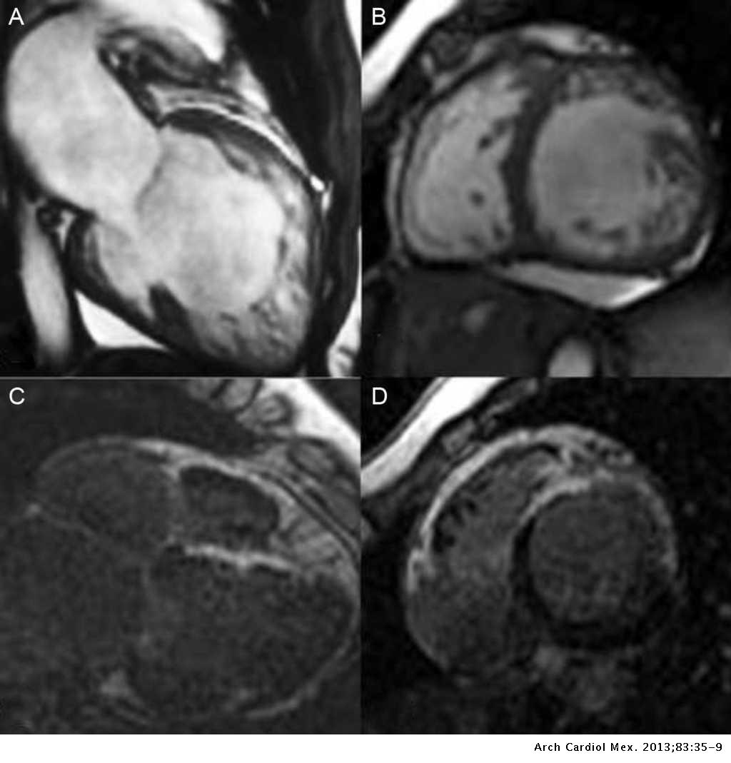
Left Ventricle Non Compaction Cardiomyopathy Different Clinical Scenarios And Magnetic Resonance Imaging Findings Archivos De Cardiologia De Mexico

Echocardiography In The Diagnosis Left Ventricular Noncompaction Topic Of Research Paper In Clinical Medicine Download Scholarly Article Pdf And Read For Free On Cyberleninka Open Science Hub
Http Medreviews Com Sites Default Files 17 02 Ricm153 8 Min Pdf
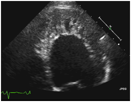
Left Ventricular Noncompaction
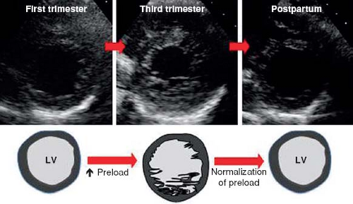
Left Ventricular Noncompaction Cardiomyopathy Thoracic Key

Isolated Left Ventricular Noncompaction Report Of A Case From Ibadan Nigeria Ogah Os Adebayo O Aje A Koya Fk Towoju O Adesina Jo Adeoye Am Adebiyi Oladapo Oo Falase Ao

Left Ventricular Noncompaction Diagnosed Following Graves Disease Habib H Hawatmeh A Rampal U Shamoon F Avicenna J Med
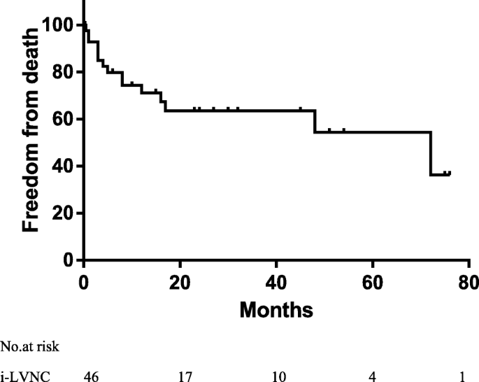
Do Children With Left Ventricular Noncompaction And A Noncompaction To Compaction Ratio 2 Have A Better Prognosis Bmc Pediatrics Full Text

Diagnostic Assessment And Therapeutic Strategies Download Table
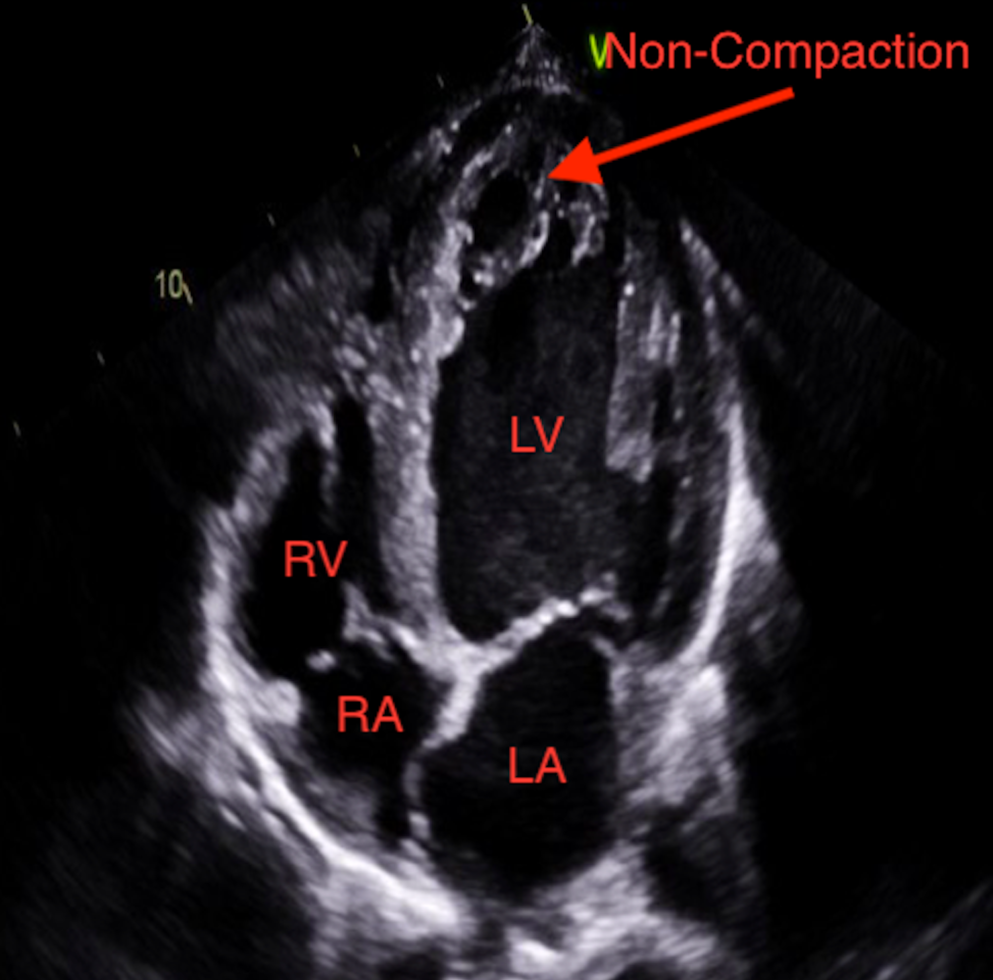
Cureus Heart Failure Secondary To Left Ventricular Non Compaction Cardiomyopathy In A 26 Year Old Male
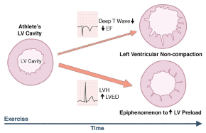
Left Ventricular Non Compaction A Review Of Literature On Clinical Status And Meta Analysis Of Diagnostic And Clinical Management Methods
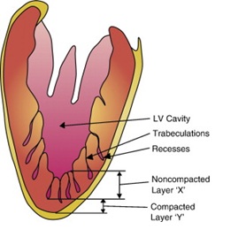
British Cardiovascular Society
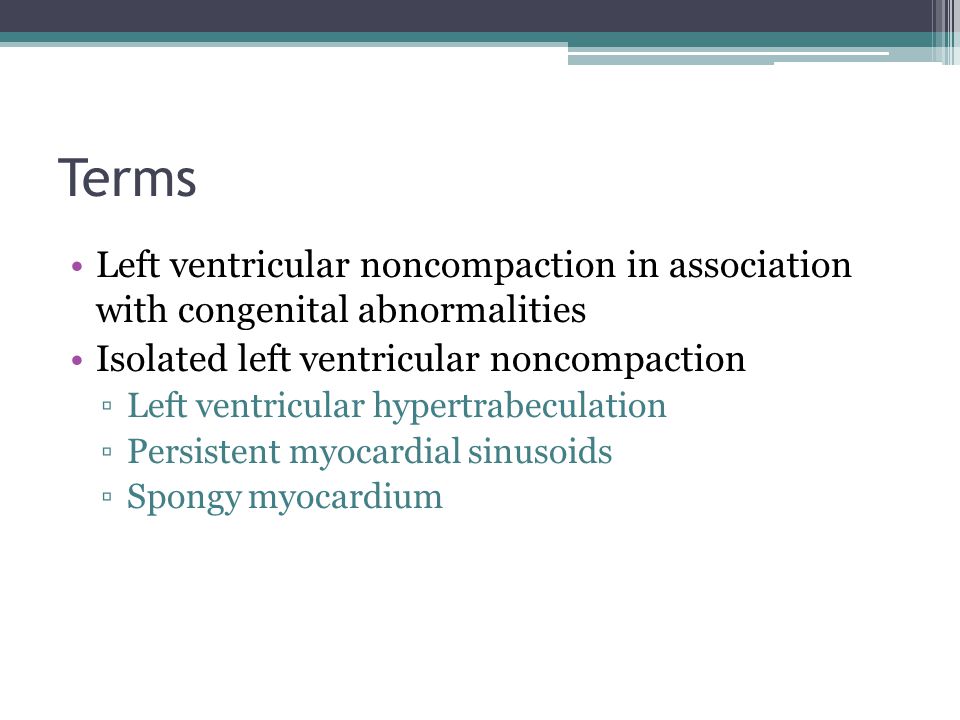
Echocardiography Conference Connie Tsao Jan 21 Ppt Video Online Download
Non Compacted Cardiomyopathy Is There A Need Of A New Cardiomyopathy

Spot The Difference Lv Trabeculation Vs Lv Noncompaction

Left Ventricular Non Compaction Cardiomyopathy Cardiomyopathy Uk
Www Onlinejacc Org Content 64 17 1840 Full Pdf
Left Ventricular Non Compaction As A Potential Source For Cryptogenic Ischemic Stroke In The Young A Case Control Study
Www Ecronicon Com Eccea Pdf Eccea 02 Pdf

Left Ventricular Noncompaction

Jpma Journal Of Pakistan Medical Association

Isolated Non Compacted Left Ventricle A Diagnostic Dilemma Chaturvedi M Singh O Agarwal A Heart India

Left Ventricular Noncompaction Intechopen

Left Ventricular Non Compaction Jacc Journal Of The American College Of Cardiology

Left Ventricle Non Compaction Cardiomyopathy Different Clinical Scenarios And Magnetic Resonance Imaging Findings Archivos De Cardiologia De Mexico
Www Sads Org Sads Media International Conference Items 17 slides Sads Lvnc 17 Aj Pdf
Undulating Clinical Course Of Noncompaction Cardiomyopathy Revista Espanola De Cardiologia English Edition

Left Ventricular Noncompaction
Multimodality Imaging Of Left Ventricular Non Compaction Cardiomyopathy Associated With Situs Inversus
Onlinelibrary Wiley Com Doi Pdf 10 1111 Echo
Left Ventricular Noncompaction


